

Streptavidin Microbubbles for Custom Cell Separation
What Is Streptavidin?
Streptavidin is a protein deriving from the Streptomyces avidinii bacterium that exhibits a high affinity for biotin. Biotin, or Vitamin B7, facilitates many biological processes involving metabolism and synthesis in the body. Streptavidin and biotin share a strong binding affinity, and the streptavidin-biotin system is resistant to breakdown due to organic solvents, denaturants, detergents, enzymes, and extremes of pH or temperature.
Streptavidin molecules are arranged as a tetramer of beta-barrels with biotin-binding sites at one end of each barrel. The streptavidin-biotin complex features a network of strong hydrogen bonds that contribute to the robust binding affinity, as well as a “lid” of β-strands that forms over the binding pockets, reducing the likelihood that biotin will dissociate from the streptavidin structure.
Applications of the Streptavidin-Biotin Complex
Biotin can couple to proteins or antibodies without degrading the way they function or the strength of the streptavidin-biotin bond. Many procedures in life sciences research rely on the bond formed between streptavidin and biotin to detect, track, and manipulate specific cells or cellular activity. For example, biotin-streptavidin flow cytometry protocol involves mixing proteins or antibodies labeled with biotin into a sample, then deploying bioconjugates containing streptavidin to capture specific cells or molecules that interact with the biotin-tagged compounds; a flow cytometer can then measure the fluorescence of the target cells or particles to analyze their physical or chemical characteristics.
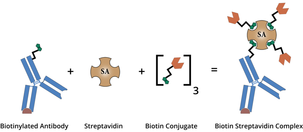
The streptavidin-biotin interaction can be utilized to identify proteins, nucleic acids, lipids, or other biomolecules within a biological sample. As well, manipulating the ways in which streptavidin and biotin bind has allowed researchers to build nanoscale structures to advance the field of bionanotechnology. Moreover, the affinity between streptavidin and biotinylated antibodies features heavily in cell sorting, separation, and isolation techniques that are essential for testing therapeutic compounds and developing patient-specific treatments.
Streptavidin Beads Protocols
Due to the prevalence of biotin in organic processes, introducing streptavidin to a biological sample allows scientists to utilize the strong streptavidin-biotin interaction to attach biomolecules to one another or target specific compounds within the body. Biotinylated streptavidin is one of the most widely used methods for identifying and manipulating bioconjugate molecules, and the streptavidin-biotin complex is often leveraged to facilitate cell sorting and separation protocols.
For example, in streptavidin beads cell sorting methods, researchers mix beads featuring a streptavidin coating into biological samples. The biotin found in various antigens, antibodies, and nucleic acids in cellular material or in biotinylated antibodies conjugated to the surface of cells quickly binds to the streptavidin coating on the beads, and both the beads and their bound targets can subsequently be removed from the sample. Separated from their sample environments, the beads and their targets can then be analyzed in biotin-streptavidin flow cytometry protocols to understand the characteristics of the captured cellular material.
Along these lines, researchers can also use streptavidin beads to eliminate unwanted cells or cellular material from a sample. After binding to the biotin molecules on the surface of target cells or cellular debris, the streptavidin beads and the material they capture can be removed from the sample, leaving behind a population of viable cells for testing or downstream analysis.
Streptavidin Magnetic Beads
One of the most common methods of using the streptavidin-biotin complex for bioconjugate tracking or cell sorting employs magnetic beads coated with streptavidin. Once the beads are mixed with a sample, they capture their biotinylated targets. Sorting or separation is then achieved via applying a magnetic field to the sample, which pulls the beads and their bound targets away from the remaining sample population.
As with all magnetic bead-based procedures, streptavidin magnetic beads protocols rely on a variety of magnetic apparatuses specific to the assays being performed to generate the magnetic fields required. Laboratory magnetic columns are costly in terms of both resources and training, which may preclude small labs or biotech startups from employing magnetic bead-based methods. Finally, while streptavidin magnetic beads manufacturers claim that their products are gentle enough to preserve the viability of fragile cells, subjecting samples to magnetic forces can inevitably degrade the health of cell populations and lead to contamination of samples from lysing.
Streptavidin Microbubbles
Microbubbles used in cell sorting and separation protocols are small, air-filled microspheres that are functionalized to bind to specific epitopes on the surface of target cells or other analytes. As with magnetic beads, the microbubbles are mixed with a biological sample in order to capture their targets; unlike magnetic beads, microbubbles rely on simple buoyancy principles rather than magnetic fields to separate unwanted cells or compounds from healthy populations. When microbubbles are mixed into the sample, they bind to their targets and then gently float to the surface of the sample container for collection or removal.
Akadeum Life Sciences’ patented Buoyancy Activated Cell Sorting [BACS] with microbubble technology is both efficient and versatile, allowing labs to use streptavidin for custom cell separation. Microbubbles coated with streptavidin are capable of targeting, capturing, and sorting cells, bacteria, viruses, or molecular analytes. . Akadeum’s Streptavidin Microbubbles offer the flexibility to create a customized isolation workflow to suit specialized research and development requirements. With Akadeum’s breakthrough BACS microbubble technology, a cell population of interest can be quickly and easily enriched even from complex samples. The ability to achieve highly targeted depletion that maintains the health and physiology of cells of interest is sure to yield better and more relevant data from downstream applications.
Cell Isolation with Akadeum’s BACS Kits
Akadeum Life Sciences developed BACS microbubble technology to disrupt industry standard cell sorting techniques with a method that is gentle, effective, and fast enough to conduct cell sorting protocols at the scale of even the most fragile cell populations. Akadeum’s cell sorting kits are affordable, easy-to-use, and require no additional equipment—perfect for small labs, startups, and research institutions alike looking to optimize their cell sorting procedures.
Akadeum’s range of products includes T cell sorting and B Cell sorting kits tailored for sorting either mouse or human immune cells, as well as Red Blood Cell Depletion kits designed for removing red blood cells [RBCs] from samples suffering from significant RBC contamination.
Akadeum’s microbubble kits function via the simple physics of buoyancy—the functionalized microbubbles bind to their target analytes, and then gently lift their targets to the surface of the sample container for removal without disturbing or compromising the health of the remaining cell population. With Akadeum’s BACS methodology, labs can quickly remove unwanted biological material from samples and maintain the integrity of remaining cells, ensuring high-purity samples for analysis and downstream applications.
Shop Streptavidin Microbubbles
Schedule a Call with an Akadeum Scientist to discuss your application.