

Leukopak Processing: Pan T Cell Isolation From Leukopaks | Akadeum
Updated on Mar 28, 2025 Share

Processing Leukopaks for T Cells
What Is a Leukopak?
A leukopak is an enriched apheresis product collected via leukapheresis and made up of white blood cells, plasma, platelets, and red blood cells. Leukapheresis is a specialized application of apheresis protocol, which is designed to extract leukocytes—white blood cells—and return the remaining cells and plasma to the donor. The product of this procedure is a highly concentrated solution of blood components from a single donor.
Leukopak Cell Count
A full leukopak volume will contain between six to ten billion cells. A leukopak is made up of roughly 50% T cells, 20% monocytes, 20% B cells, 20% NK cells, and 6% granulocytes and hematocrit. The white blood cell count of a leukopak is over six times greater than in whole blood, and 20 times greater in the concentration of PBMCs.
Leukopak vs. PBMC
Leukopaks can be processed into human peripheral blood mononuclear cells (PBMCs) by washing away residual red blood cells and plasma, leaving a high concentration of mononuclear cell components. PBMCs are formed from stem cells within the bone marrow and released into the blood.
PBMCs play an integral role in the body’s first line of immune defense, as they are constantly circulating through the bloodstream looking for infection. The majority of a PBMC enriched sample contains lymphocytes—the most common white blood cell—monocytes, and dendritic cells. This increase in concentration is preferable to researchers in need of a large number of white blood cells from a single donor, providing the ability to perform multiple experiments using one leukopak.
Many different fields of research use leukopaks, including immunology, cell therapy, and drug and vaccine creation.
Leukopak Processing
Leukopaks can be further processed into their cellular components—such as basophil isolation, PBMCs, or T cells—using various cell separation techniques. These techniques involve prepping a leukopak by washing its contents, removing residual red blood cells and platelets, and performing additional cell separation procedures to target the intended cells.
This process can be arduous and risky. If done incorrectly, the final result could be an unfeasible viable cell count yield for future experimentation. There are multiple techniques available for cell separation, so choosing the right one for your research is important.
Pan T Cell Isolation Protocol
Isolating Pan T cells from leukopaks can be a lengthy and expensive process, one that involves crucial cell separation steps, multiple magnetic columns, and small volume maneuvering. The protocol begins with the isolation of PBMCs via density gradient centrifugation to obtain a buffy coat. By this step, the enriched PBMC product is extremely valuable, and using a reliable separation technique to isolate T cells is imperative to optimizing the final yield.
The T cells are then either positively or negatively selected via chosen isolation methods (magnetic, buoyancy, or fluorescence). In positive selection, the cells of interest are directly selected and separated from the solution. Inversely, negative selection targets unwanted cells, leaving only the cells of interest behind in the solution. Although negative selection can result in lower yields, it remains the preferred isolation technique because positive selection can trigger T cell activation.
Required Materials
- Leukopak prepped to be enriched with PBMCs
- T cell Isolation Kit (magnetic, buoyancy, fluorescence-activated, etc.)
- Sterile containers
- 50mL conical tubes
- Separation buffer
- Hemocytometer (for cell counting)
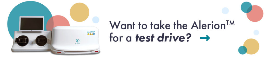
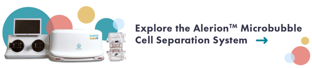
BACS™ Microbubbles
Many cell separation techniques exist, but few maintain the integrity of your desired cells the way that Akadeum’s microbubbles for cell therapy do. Using antibody labels, our Human T Cell Isolation Kit only targets the T cells through negative selection. By using Buoyancy Activated Cell Sorting (BACS™), the T cells will separate and the unwanted cellular components will float to the top to be either discarded or stored.
Our microbubble technology also works straight from a leukopak, meaning there is no need for pre-processing or enrichment and you can even perform the protocol within the leukopak bag itself. The T cells will self-separate within a single container, minimizing experimental error resulting from transferring liquids and changing tubes. This decrease in error rates and ease of use is what sets our BACS™ microbubble technology apart from standard magnetized approaches.
Isolation From Leukopak vs. Whole Blood
What are some of the benefits of isolating T cells from leukopaks rather than from whole blood?
- You can get a large amount of viable white blood cells from a single donor.
- You end up with a higher concentration of mononuclear cell content than in whole blood.
- Leukopaks contain peripheral blood cells which provide the first line of immune defense against disease. This makes it an excellent material for studying how the body responds to infections as well as vaccines and drugs.
Leukopak Processing for Cell Therapy
One of the major applications of leukopaks is within cell therapy research. Due to its single-donor nature, a leukopak sample is a robust and concentrated sample to perform analysis on, providing 20 times more mononuclear cell content.
Some leukopaks are specially designed for ideal ratios of blood components. Notably, leukopaks assist in studying diseases that affect the blood and our immune response—like HIV and AIDS—and provide a reliable source of cell material for researchers to accurately test the immune system.
What Is a Mobilized Leukopak?
A mobilized leukopak is specifically extracted from healthy donors who have been treated with mobilization factors intended to boost the release of progenitor cells from the bone marrow. Mobilized leukopaks have a higher concentration of stem cells and are useful in regenerative medicine, transplant therapy, and cell therapy applications.
Mobilized Apheresis
Mobilized leukopaks are obtained through a mobilized apheresis donation—a single consenting donor on a set regimen of drugs aimed to mobilize stem cells from the bone marrow into the blood to be collected via leukapheresis. These drugs include plerixafor and G-CSF (granulocyte-colony stimulating factor), both shown to activate a release of CD34+ cells into the bloodstream of the donor for collection into a mobilized leukopak via leukapheresis. By mobilizing stem cells into the bloodstream for collection, the resulting mobilized leukopak will be rich in concentrations of CD34+ cells, all from a single donor.
Akadeum Offers an Innovative Solution for Leukopak Processing
Akadeum’s BACS™ microbubble technology is a much-needed reimagination of cell separation techniques. It can save researchers precious time, resources, and funds by both reducing reactions to a single tube and being highly customizable to target Pan T cells.
The margin of error from harsh cell environments, tube switching, and pour-offs can become a thing of the past in the lab. Using Akadeum microbubbles, a time-consuming and risky preparation step can become a robust protocol with high yield and reliable delivery every time, allowing researchers to focus on the T cell studies at hand.
Considering an Akadeum BACS™ Microbubble Kit for Pan T Cell Separation for your next cell separation experiment? Contact us today.