

What Is PBMC? Human PBMC Cells, PBMC Composition, and Immune Cell Processing
Updated on May 19, 2025 Share
Peripheral Blood Mononuclear Cells
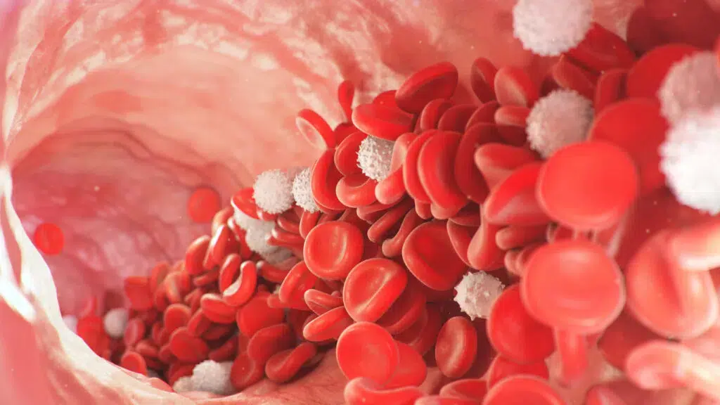
Peripheral blood mononuclear cells (PBMC) are crucial immune system cells that protect the body against harmful pathogens. Human PBMCs include T cells, B cells, NK cells, monocytes, and dendritic cells. These cells are essential in immune response and are often used in research and clinical applications due to their versatility. Located in peripheral blood, these cells act as a primary defense against infection and disease, making PBMC isolation from whole blood samples a common practice in medical studies. Read on to learn more about the importance and functions of PBMCs.
Where Do PBMCs Come From?
PBMCs are developed in the bone marrow from hematopoietic stem cells (HSCs). HSCs are the building blocks of all blood cells in the immune system. As they undergo a process called hematopoiesis and differentiate to different cells, they develop two different lineages — myeloid and lymphoid. PBMCs include certain myeloid and lymphoid cells, particularly those that have a single round nucleus.
As HSCs develop into different lymphocytes, dendritic cells, and monocytes, they help bolster the adaptive and innate immune systems. From the bone marrow to the peripheral bloodstream, mature PBMCs will assist the body in defending against bacteria, viruses, and diseases by destroying foreign substances and tumor cells.
PBMC Cell Types:
The three main cell categories present in PBMC composition include:
- Lymphocytes
- Monocytes
- Dendritic Cells
Each of these PBMC cell types is responsible for carrying out a specific function.
1. Lymphocytes
The majority of an enriched human PBMC sample is composed of lymphocytes — about 70-90 percent. Lymphocytes are the most abundant white blood cells (WBCs) because there are multiple different types that all serve a significant role in responding to diseases and infections. In a human PBMC population, lymphocytes are broken down into the following categories:
- The total number of T cells (CD3+) – Roughly 45-70 percent of the lymphocytes in a sample will be naive, resting CD3+ T cells. These lymphocytes have yet to be activated through antigen recognition and travel freely through the bloodstream until needed. These cells could also include memory cells, which are already primed to combat a specific antigen and will reactivate upon exposure. Memory T cells help individuals to build up long term immunities while naïve T cells create the adaptive immune system to battle new harmful substances.
- CD4+ helper T cells – When CD3+ T cells are activated, they primarily develop into CD4+ or CD8+ T cells. CD4+ T cells are called helper T cells because their primary function is to activate an immune response. They use receptors to bind to antigen-presenting cells (APCs) and release chemicals to signal the location of invasive cells and mark them for elimination. Helper T cells make up between 25-60 percent of total activated CD3+ cells.
- CD8+ killer T cells – The other differentiated T cell is called a CD8+ killer T cell. Sometimes called cytotoxic T cells, these specialized lymphocytes are responsible for producing the antibodies that seek out and destroy invasive cells. About 5-30 percent of activated CD3+ cells become killer T cells.
- B cells – Similar to T cells, most B cells exist in the bloodstream as naive or memory cells. Once activated by an antigen, they differentiate into plasma cells that can target freely circulating antigens. By secreting antibodies into extracellular space, they can track down and destroy harmful cells that T cells cannot reach. B cells make up around 5-15 percent of the total lymphocyte population.
- Natural killer (NK) cells – A small portion of lymphocytes, roughly 5-10 percent, are natural killer cells. These cells are viewed as the human body’s innate front-line defense system. While T and B cells need to be activated by antigens to perform their immune functions, NK cells carry out their roles without the need for antigen activation. They are always active in defending the body from tumor activity.
2. Monocytes
Monocytes make up a significantly smaller portion of the human PBMC sample than lymphocytes — roughly 10-30 percent. These are the largest type of white blood cells and are typically found in peripheral tissue. When monocytes are stimulated, they can differentiate into macrophages or dendritic cells. Macrophages help shape the adaptive immune system by disposing of unnecessary or dead cells.
Unless they turn into memory cells, effector cells (CD4+ and CD8+) are no longer useful to the immune response after they carry out their function. Similarly, after an invasive virus, parasite, or otherwise harmful cell is destroyed by killer T cells, killer B cells, or NK cells, its cellular debris must be cleaned from the bloodstream. Macrophages consume and dispose of extracellular waste, while dendritic cells use cell fragments to further bolster the body’s immune response.
3. Dendritic Cells
The third type of PBMC is the dendritic cell. Dendritic cells are highly specialized APCs that are capable of engulfing an antigen completely, then presenting a small part of the same antigen to the immune system. When the dendritic cell presents the APC fragment, T and B lymphocytes become activated and launch their response against the cell population presented.
Dendritic cells are a type of monocyte that only make up 1/100 of the total PBMC composition percentage. Though low in concentration, these cells are crucial in identifying unknown substances and communicating with lymphocytes.
PBMC Application
Each of these cell populations plays an integral role in building and maintaining an effective immune response. For this reason, medical researchers can use them to study immune cell behavior when exposed to harmful pathogens, disease progression in the human body, and factors that affect long-term immunity. PBMCs are used to study a multitude of areas, including (but not limited to):
- Infectious diseases
- Vaccine development
- Immunology
- Primary cancers of the blood and blood-related organs
- Personalized medicine
- Toxicology
- Transplant therapy
- Disease modeling
- Biomarker identification
Isolated PBMCs have become a critical part in many subdivisions of medical research.
On top of the value they provide for scientific advancements, they are also being used for medical applications on patients who may need help that can’t be provided through traditional methods. This includes the use of genome editing technology to engineer immune cells for personalized medicine and cancer treatment. Scientists have begun using gene and protein therapy to treat certain patients who have exhausted other options, but to carry out those procedures they require a sufficient population of PBMC T cells to alter and reintroduce into the sick individual.
The need for healthy isolated PBMC samples has dramatically increased, and many separation techniques can’t keep up with the current demand or have inherent limitations that make PBMC isolation difficult to scale.
Isolating PBMCs
There are two primary methods for separating peripheral blood mononuclear cells from whole blood: density gradient centrifugation and leukapheresis. Each of these methods has its own advantages and disadvantages.
Density gradient centrifugation separates cell populations throughout a density gradient medium. The sample is spun in a centrifuge with a substance of a particular density that helps sort lymphocytes, plasma, and RBCs. After the whole blood is centrifuged, the PBMC layer will appear as a thin white layer below the plasma.
Although the process is relatively cheap and simple, density gradient centrifugation is not very efficient. For large-scale research studies or trials, it may be more beneficial to use leukapheresis. Leukapheresis can supply 14 times the number of PBMCs from a single donor than centrifugation.
Leukapheresis requires a machine that separates the PBMC fraction of whole blood from other components with high-speed centrifugation, using a machine that processes this collection in real-time during the blood draw. As the leukapheresis device separates the various components from the whole blood, it collects and isolates the leukocytes and returns the other cells such as plasma, RBCs, and granulocytes back to the whole blood donor. PBMCs can also be isolated by using cell sorting techniques such as magnetic-, fluorescent-, and buoyancy-activated cell sorting.
PBMC Characterization
After PBMC processing, the mononuclear immune cell sample is placed into a flow cytometer. A flow cytometer is a device that identifies and sorts different cell populations based on physical characteristics such as complexity, size, and biomarker expression through a process called flow cytometry.
Regardless of the method used for peripheral blood mononuclear cell isolation, the sample will likely be contaminated with residual red blood cells (RBCs) and granulocytes. In comparison to leukocytes, granulocytes are much larger and more intricate, and RBCs are smaller and less intricate. Flow cytometry is capable of differentiating these contaminants from PBMCs. With an extra biomarker, a flow cytometer can also identify and sort healthy or damaged WBCs for further purification.
Evolving medical technology also enables the flow cytometer to provide further insight into a PBMC fraction with in-depth immunophenotyping. Essentially, these machines can help us continue to discover more about an individual based on the concentration and condition of cells located in a PBMC sample. This includes information on cellular function and how far along a disease may have progressed in a patient’s body.
Akadeum’s Technology
PBMC separation can be an arduous and costly process. At a time when isolated leukocytes are critical to advancing medical treatment and personalized medicine, it’s important to be as efficient as possible. Akadeum Life Sciences has developed an innovative technology that helps to reduce the resources necessary for cell separation.
Buoyancy activated cell sorting (BACS) is a technique that harnesses the buoyant properties of microbubbles to float target cells. By attaching antibodies to the bubbles and binding those antibodies with antigen receptors on target cell populations, the bubbles will gently carry target cells to the top of the sample container. This is typically done through negative selection, meaning the bubbles lift unwanted substances to the top of the container for removal, leaving the enriched cells of interest pelleted at the bottom for easy collection.
The process of BACS doesn’t require expensive equipment or trained personnel and can be done directly in the sample container. While the bubbles can be used for cell isolation on their own, they also work with other techniques to further purify samples, delivering a highly enriched sample for downstream applications.
Microbubbles and PBMC Isolation
BACS can be paired with any isolation method to further enrich a PBMC sample after centrifugation or leukapheresis. Akadeum’s Human RBC Depletion microbubble kit can remove residual RBC contamination from a PBMC sample in an exceptionally gentle, 10-minute workflow that eliminates the need to expose delicate immune cells to harsh chemicals like lysis buffers. If you have contaminants in your peripheral blood mononuclear immune cell population, Akadeum’s microbubbles could be the solution to your sample preparation headaches. They work quickly and gently to provide high throughput while preserving cell health and physiology. For more information on RBC Depletion in PBMCs, you can download our app, or get started with one of our Human RBC Depletion Kits today.