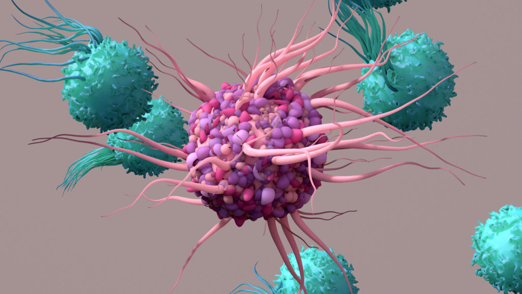
Human T Cell Activation
T cell activation is a complex interaction of molecular signals and cellular responses that trigger the immune system. This crucial process in the immune system’s defense against infection and disease allows T cells to effectively identify and respond to foreign antigens.
Understanding T cell activation is vital for developing targeted immunotherapies and vaccines. T cell activation also plays a key role in maintaining the immune system’s equilibrium, helping to prevent overreactions and autoimmune disorders. Dive deeper into the mechanisms and significance of T cell activation in the following sections.

The Vital Role of T Cells in Immune Dynamics
The immune system defends the human body against infections and diseases through a complex assembly of cells, tissues, and organs. T cells play a vital role in orchestrating immunity. They are not just defenders; they are also strategists who modulate responses to different threats.
As essential components of the adaptive immune system, T cells are known for their ability to recall and mount effective responses against previously encountered pathogens. This immunological memory enables the body to swiftly counter familiar infections.
T cells collaborate with other immune cells like B cells and macrophages, recognizing specific antigens for a targeted response. They play a key role in preventing autoimmune diseases by distinguishing between self and non-self entities.

Where Does Activation Occur?
Like most lymphocyte activation, T cell activation occurs in the secondary lymphoid organs when the cell locates its predetermined antigen. Lymphocytes constantly circulate through the secondary lymphoid organs: the spleen, lymph nodes, tonsils, and unique lymphoid structures within mucosal tissues.
Why Is Human T Cell Activation Necessary?
Human T cell activation is imperative to successful adaptive immune response and allows for the resilience of human immunity to developing diseases. Human T cells are, therefore, an essential subject for study and can help us further understand the mechanisms underlying effective immunity.
Activation Steps
The activation of T cells is a complex process that involves several critical steps. Each step has a specific role in ensuring T cells are correctly activated to combat pathogens. This sequence of events is vital to the immune system’s ability to initiate an effective response and involves a combination of molecular recognition, signaling, and cellular response.
Step 1: Antigen Recognition
T cell activation begins with antigen recognition. T cells, using their T cell receptors (TCRs), specifically recognize antigens presented on major histocompatibility complex (MHC) molecules by antigen-presenting cells (APCs) like macrophages.
MHC molecules are crucial for immune function, displaying antigen-derived peptide fragments for T cell surveillance. This recognition ensures that T cells accurately identify and respond to specific antigens, a fundamental step for targeted immune response.
Step 2: Costimulatory Signals
T cells require a second signal from costimulatory molecules on APCs following antigen recognition. These molecules, including CD28 and CD3, are essential for full activation. CD28 amplifies the TCR signals, while CD3 transduces these signals into the cell, leading to T cell activation. This step ensures that T cells are fully equipped and committed to responding to the antigen.
Step 3: Cytokine Secretion and Response
Once activated, T cells secrete cytokines, which are critical for modulating the immune response. These proteins have various effects, from stimulating further T cell activation to controlling inflammation. This phase is pivotal in dictating the scope and scale of the immune response, fine-tuning the body’s defense against specific microbes and diseases.
Cytokines play a crucial role in eliminating infections by inducing collective cell death. This makes them an important tool in cancer research and fighting cancer within the body. However, it’s important to control cytokine activity during adoptive cell therapy to prevent cytokine release syndrome in patients. This syndrome can rapidly damage healthy tissues, hence the imperative need for its prevention.
Step 4: Proliferation and Differentiation
During the final stage of the immune response, T cells undergo proliferation and differentiation. The activated T cells multiply rapidly and transform into various types, such as helper T cells or cytotoxic T cells, each with a specialized function in the immune system. This phase is essential for amplifying the immune response and ensuring a robust defense against diseases.
Throughout this process, signaling pathways play a crucial role. They transmit information from the cell surface to the nucleus, guiding the T cell’s response. These pathways are intricate networks involving various molecules and enzymes, each carefully regulated to ensure a precise and appropriate response.
Understanding these steps in T cell activation not only provides insights into the fundamental workings of the immune system but also highlights potential targets for therapeutic interventions in diseases where the immune response is compromised or overactive.
Exploring the Diversity and Activation of T Cell Types
T cell diversity, encompassing various types with unique functions and activation mechanisms, is vital for the body’s response to different pathogens and conditions. Each T cell type is activated through interactions with antigens presented by APCs, leading to distinct pathways and roles.
- Naive T cells: As precursors to effector and memory T cells, naive T cells circulate in the bloodstream, each bearing a unique T cell receptor ready for activation. Most will remain inactive, only activating upon encountering their specific antigen. Following activation, they proliferate and differentiate into the necessary T cell types. Post-infection, most active T cells die, leaving behind memory T cells for potential future exposures, thus contributing to the development of new immunities.
- Cytotoxic T cells (CD8+): During an infection, cytotoxic T cells are activated by recognizing antigens presented by MHC class I molecules. Upon activation, they target and destroy infected or abnormal cells.
- Helper T cells (CD4+): Once activated, helper T cells play several roles in the body’s inflammatory response. They communicate with other cells through cytokines and effector molecules and can directly activate cytotoxic T cells and B cells for antibody production.
- Regulatory T cells (Tregs): Tregs are essential for maintaining immune tolerance and preventing autoimmune reactions. They regulate other immune cell activity to avoid damaging the body’s tissues.
- Memory T cells: These long-lived cells retain the memory of past infections. Upon re-exposure to the same antigen, they rapidly proliferate and mount a strong response, underpinning immunological memory.
Deciphering the Signaling Pathways in T Cell Activation
Signaling pathways play a crucial role in activating T cells, controlling the entire sequence of events from the recognition of antigens to the final response. When the TCR recognizes the antigen-MHC complex, intracellular signals are initiated, which activate kinases and trigger gene transcription for T cell proliferation, differentiation, and cytokine production. These activation signals are further boosted by costimulatory signals, especially from CD28.
Understanding these signaling pathways is essential for comprehending immune system responses and identifying therapeutic targets. Disruptions in these pathways can result in immune deficiencies or autoimmune diseases, while targeted manipulation can offer new treatments in cancer immunotherapy and immune-related disorders.
Implications of T Cell Activation Research in Medicine
T cell activation research is revolutionizing clinical medicine, especially in immunotherapy for cancer. Scientists have developed therapies to enhance the immune system’s ability to target and destroy cancer cells by manipulating T cell responses. TCR and CAR-T therapy are two primary examples.
Understanding how T cells are activated by specific antigens is important for vaccine development. It is also crucial in treating autoimmune diseases, which occur when the immune system mistakenly attacks the body’s own cells. By comprehending the mechanisms of T cell activation, medical professionals can develop therapies to regulate this response, thus preventing harmful autoimmune reactions.
The ongoing research in T cell activation is expanding our knowledge of the immune system and creating new opportunities for treating various diseases. This highlights the significance of this field in immunology and medical science.
Can T Cells Be Artificially Activated?
Yes, there are several in-laboratory approaches for T cell activation. This practice provides a controlled method for studying T cell biology or prime therapeutic T cell populations. This can be facilitated through the innovation of APC-mimetic scaffolds (APC- ms), which artificially present chemical signals to T cells to mediate activation and rapid T cell expansion.
More often, however, activation is achieved in vitro through the use of soluble or particle-bound antibodies against CD3 and CD28 that lead to generating activation signals.

Akadeum’s Advanced Approaches to T Cell Activation
Akadeum Life Sciences is at the forefront of providing innovative solutions for T cell activation research and applications. Our cutting-edge T Cell Activation and Expansion kit facilitates the separation and activation of T cells, enabling researchers to study these critical immune cells more effectively. Akadeum’s microbubble technology offers a gentle and efficient method for isolating activated T cells, ensuring their functionality and health preservation.
In addition, the Human T Cell (GMP grade) and peripheral blood mononuclear cell (PBMC) Isolation kits deliver high-purity populations of activated T cells when collected during T cell expansion. Our kits separate large numbers of T cells for any downstream applications while significantly reducing sort times. The gentle nature of the negative selection protocol is ideal for fragile cell populations, helping defend the isolated cells’ health and physiology.
Advanced techniques from Akadeum allow for high-purity populations of activated T cells, crucial for immunology research and developing new therapies.
Akadeum’s T Cell Activation Protocol
Embracing the Complexity and Importance of T Cell Activation
The study of T cell activation is not only an important component of immunology, but it also holds the key to discovering new medical breakthroughs. As we delve deeper into this complex process, we come closer to finding revolutionary treatments for various diseases. Akadeum is leading the way in this research with its innovative products, providing researchers and clinicians with the necessary tools to pioneer these advancements.
Join us in shaping the future of healthcare. Reach out to discover how our cutting-edge solutions can power your research and clinical breakthroughs, marking a new era in advancing immunological science.


