

What is Buffy Coat in Blood? Buffy Coat Preparation and Buffy Coat Cell Extraction
Updated on May 16, 2025 Share
What Is a Buffy Coat?
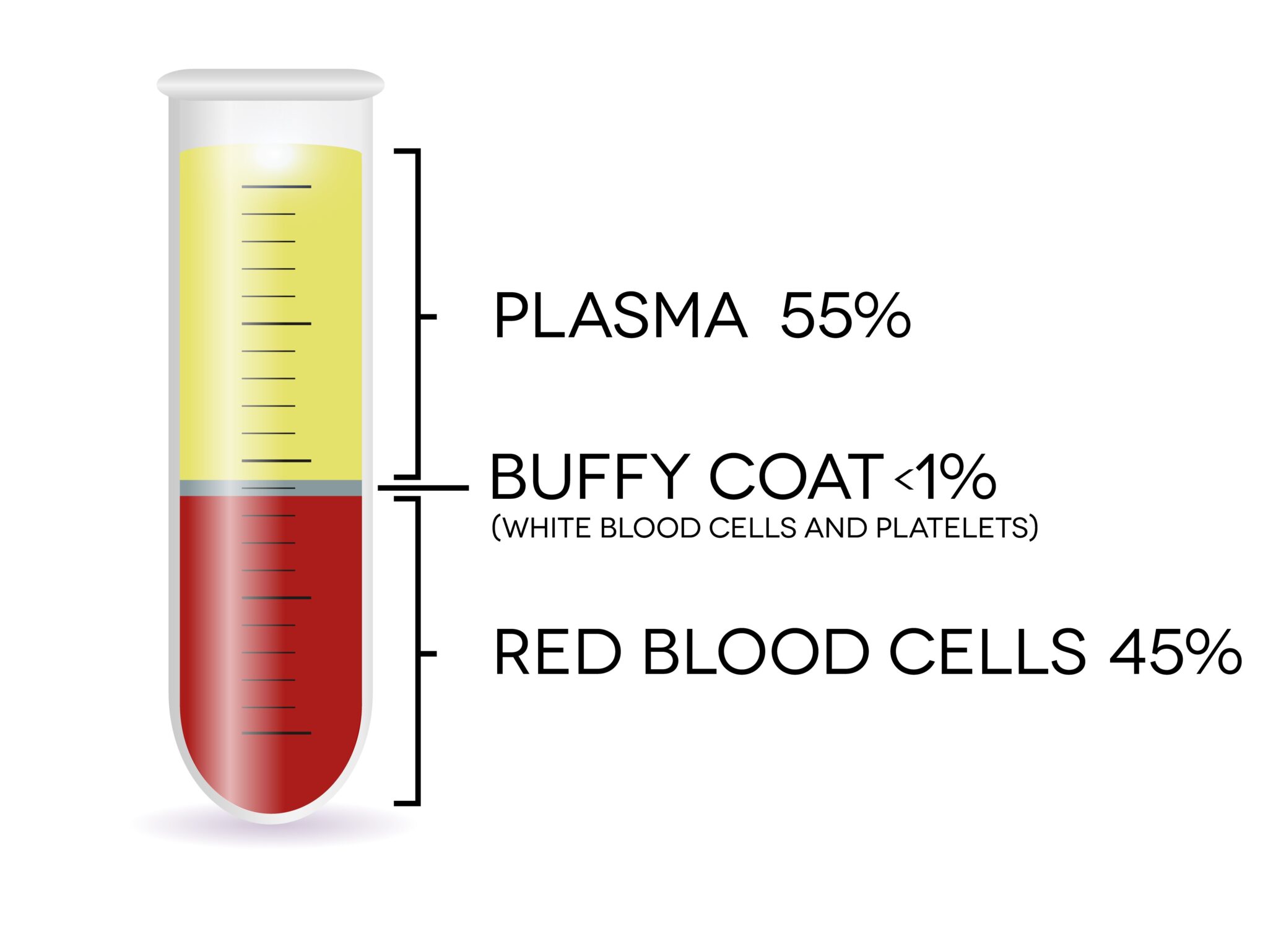
A buffy coat is a mix of lymphocytes, monocytes, granulocytes, and platelets, isolated from plasma and RBCs by centrifugation. In a sample of peripheral whole blood, less than 1% of the cells are white blood cells (WBCs) and platelets. When researchers put the sample through a centrifuge, a machine that spins the blood, those WBCs and platelets combine to form their own layer suspended between the red blood cells (RBCs) and supernatant plasma. This thin layer is called a buffy coat because of its color (yellowish to brownish).
Leukocytes, or white blood cells that help the body fight off infection, are 10-20X more concentrated in the buffy coat than the total sample. This can be useful for researchers doing work with toxicology or for patients with dysfunctional WBCs or platelets.
Buffy Coat Cells
A buffy coat consists mainly of four types of cells:
- Lymphocytes – A type of WBC that plays a key role in the human immune system by producing antibodies. Lymphocytes can be further divided into T cells and B cells, along with natural killer cells.
- Monocytes – Another type of WBC that helps in the immune response by isolating and digesting harmful microorganisms. Monocytes can differentiate into macrophages or dentric cells.
- Granulocytes – A form of immune cell that can be further divided into basophils, eosinophils, and neutrophils, which secrete chemicals, attack parasites, and kill bacteria/fungi respectively.
- Platelets – Also called thrombocytes, these cells are not RBCs or WBCs. They are small colorless cell fragments that migrate around the body to form clots and prevent bleeding where necessary.
The cells that can be found in a buffy coat are essential to protecting humans from disease and healing from wounds. The medical community can benefit from studying the behavior of these cells when exposed to other pathogens.
Importance of a Buffy Coat
White blood cells are the building blocks of the human immune system. Different types work together to identify foreign substances, eliminate threats to the body, and build a sustainable defense system against future infections. Platelets are the first responders of the human body, rushing to injured areas and clotting blood flow through adhesion. Both WBCs and platelets are present and prevalent in the buffy coat. The high concentration of WBCs and platelets make the buffy coat a critical bio-fluid in the medical research field, enabling academic professionals to conduct experiments and study these essential cells and how they work in the body—which in turn can shape the direction of medicine and effective patient treatment.
Beyond its implications in research, the buffy coat also has a practical application in testing for certain diseases and treating patients. Because key immune cells concentrate in this layer, the buffy coat can be used diagnostically when looking for the presence of harmful diseases such as malaria. The platelets can also be further purified and used therapeutically to increase counts in patients who may have low platelet levels. Blood banks often separate samples of whole blood to isolate the buffy coat and store it for therapeutic use in individuals who could need this type of therapy to treat their conditions.
Buffy Coat vs. PBMC
The term buffy coat is often used interchangeably with PBMC, however, there are minor differences in cell composition that can make a large difference. PBMCs, on the other hand, are individual fragmented lymphocytes and monocytes that separate from the rest of the whole blood sample through a process called density-gradient centrifugation.
This process involves an additional density gradient that separates sample particles further based on their size and density. By separating out granulocytes and platelets and creating a barrier between the WBCs and the RBCs, the sample is comprised of a more concentrated sample of monocytes and lymphocytes that can then be used for research or further isolated for specific downstream applications.
How to Prepare a Buffy Coat
Although the contents of a buffy coat already exist within a whole blood sample, they are not aggregated until centrifugation occurs. Before centrifugation, researchers should take a variety of precautions to ensure the extraction process is successful.
The first step is to collect the whole blood sample using the correct tube – cell-free DNA collection tubes like those from STRECK, or tubes containing an anticoagulant like EDTA are two popular options. STREK tubes are commonly used for research looking to isolate DNA from blood, while EDTA tubes contain an EDTA anticoagulant used for processing whole blood samples without clotting, allowing for centrifugation into the different layers—including the buffy coat layer.
It is important to remember that a whole blood sample could contain pathogens, so it’s critical that these samples be handled with standard precautions as if capable of transmitting infectious diseases. After the necessary preparations, it’s time to extract the buffy coat from the sample of whole blood.
Buffy Coat Extraction
The prepared whole blood sample is placed into a centrifuge to fractionate the buffy coat and separate it from the plasma and RBC. After the centrifugation process is complete, there will be a thin layer between the RBC and plasma that makes up around 1% of the sorted sample—the buffy coat. Using a small pipette, the experimenter collects the buffy coat and moves it to a separate container.
PBMC isolation often involves particle separation by a density gradient in the sorted sample, meaning the buffy coat is in direct contact with isolated RBCs. Due to the size and abundance of the RBCs, using a manual extraction method like pipetting makes it extremely likely contaminating RBCs are included in the buffy coat sample (this is why buffy coats are referred to as “pink” prior to further processing). To further enrich the buffy coat, researchers will often pair centrifugation with another method of cell separation, like BACS, to remove the residual RBC contamination.
Akadeum’s Technology
BACS, or buoyancy activated cell sorting, is an innovative technology developed by Akadeum Life Sciences that uses functionalized microbubbles to isolate target cells. By attaching antibodies to the microbubbles that are specific to antigen-presenting cells in the sample, BACS can effectively target a specific type of cell and float it to the top for collection or removal. Traditional blood separation techniques require expensive equipment, long processing times, and cause unnecessary cell death. BACS, on the other hand, enables a simplified workflow that doesn’t require additional equipment and allows for sample enrichment that is exceptionally gentle on cells.
Microbubbles and Buffy Coats
Akadeum’s Human RBC depletion kit removes over 97% of residual RBCs from a sample in a fast and easy 3-step, 10-minute workflow that can be processed directly in the sample container. This method is fast, relatively inexpensive, and doesn’t expose delicate immune cells to harsh chemicals like lysis buffers, or external forces like those from rare earth magnets. Akadeum’s Red Blood Cell depletion protocol can be used to effectively and efficiently further enrich the buffy coat layer and prepare the cells for downstream applications while maintaining WBC and platelet health and physiology. For more information, you can download our app note: Removal of Red Blood Cells from Human Peripheral Blood Mononuclear Cell Samples Using Akadeum Human Red Blood Cell Depletion Microbubbles, or to experience the microbubble difference for yourself, you can get started with our Human RBC Depletion products today.