Necrosis Vs. Apoptosis: Processes, Necrotic Cell Death, & Apoptosis Steps
Updated on May 16, 2025 Share
Necrosis and apoptosis are the two main types of cell death in the body. Necrosis is often a result of injury leading to uncontrolled cell death, while apoptosis is a programmed and orderly process. Each of these involves a unique process and has different effects on the rest of the body. Understanding these differences is crucial for researchers and clinicians in developing treatments for various diseases. Read on to delve deeper into the mechanisms and implications of necrosis and apoptosis.

What is Cell Apoptosis?
Programmed cell death is called apoptosis, a regular growth, and development mechanism within an organism. This process is also known as “cellular suicide” because the cell takes part in its own death.
Just like multicellular organisms, each cell in the body has a lifespan. Some cells live longer than others, but ultimately, they must die to be replaced by healthy cells. Apoptosis helps to maintain the balance of cell multiplication. If cells constantly reproduce without apoptosis, unregulated cell growth will result in tumors and other issues.
What Causes Apoptosis?
Apoptosis is known as programmed cell death because it’s typically caused by self-generated signals within a cell. It is a natural part of the cell cycle initiated by mitosis in cell reproduction. This process is mediated by caspases, enzymes that exist in all cells, and cleave specific proteins to begin the process of cell death.
There are also other stimuli that can induce apoptosis. Stimuli such as hypoxia, certain immune reactions, high temperature, and exposure to certain chemicals can cause the body to signal apoptosis in cells. In moderation, apoptosis is used to keep the body healthy. If it occurs too quickly or slowly, however, it can cause a variety of problems. If apoptosis is happening at a decreased rate an individual may experience unregulated cell growth. If it happens too quickly, they may be experiencing an autoimmune disease such as AIDS or Crohn’s disease.
Apoptosis Steps
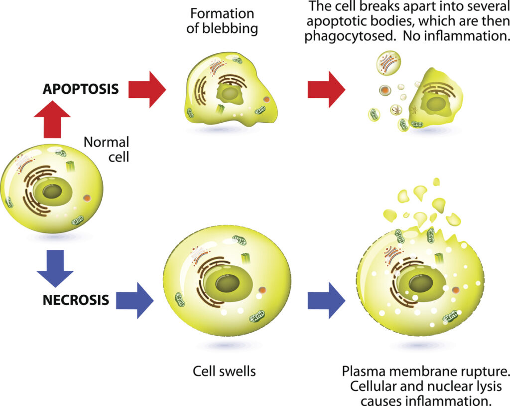
Cell Apoptosis happens in a gentle and controlled way that does not negatively affect surrounding cells. A cell will release moisture to dry itself out and condense until fragmentation occurs. There are no morphological changes because the cell does not release into the extracellular environment. Small vesicles called apoptotic bodies are formed to transport the cell contents elsewhere. This allows cells to die gently and not cause any inflammation.
Apoptosis is signaled by chromatin condensation in the nucleus of a cell. This helps scientists determine whether a cell is dying of natural or unnatural causes.

What is Cell Necrosis?
Another form of cell death called necrosis occurs in cells that are exposed to extreme conditions. Outside of normal conditions, cells may experience damage to their internal environment. Tissues may begin to deteriorate harshly and rapidly. Therefore, necrosis is typically defined as accidental cell death.
There are no vesicles formed during necrosis, which means cellular content is released into the surrounding area. This extracellular debris can have negative effects on adjacent cells.
Causes of Necrotic Cell Death
Necrosis occurs because of external factors influencing the physiology of cells in the body. Examples of necrotic cell death causes include the following:
- Bacterial infection – Bacteria are microscopic organisms that can cause infections when they enter the body through an airway or open wound. They can be responsible for a variety of illnesses that are linked to the unplanned death of cells.
- Fungal infection – Also called mycosis, occurs when any fungus invades human tissues. This could cause diseases of the skin or internal systems resulting in unprogrammed cell death.
- Pancreatitis – The inflammation of the pancreas, a gland that helps regulate hormones and digestion in the body.
- Denaturation of proteins – Proteins are responsible for nearly every process in the human body. Denaturation occurs when weak bonds in the proteins break down, damaging the cells’ ability to function properly.
While some recent studies have begun to find that necrosis may sometimes be natural, it is primarily used to describe death that occurs as a result of something unnatural.
Apoptosis Vs. Necrosis
While both necrosis and apoptosis are mechanisms involved in multicellular organism cell death, there are multiple ways in which they can be differentiated. Apoptosis is viewed as a naturally occurring process while necrosis is a pathological process. Pathological processes are caused by toxins, infections, or traumas, and are often unregulated. Apoptosis is both regulated and timely, making it predictable and healthy for the host.
The difference between apoptosis and necrosis can also be seen in the following factors:
- Process – Apoptosis involves the shrinking of cytoplasm, resulting in the condensation of the nucleus. Necrosis happens when cytoplasm and mitochondria swell up to cause cell lysis, or a rupture in the cell membrane.
- Membrane Integrity – A hallmark trait of apoptosis is blebbing. Blebbing occurs when the cytoskeleton of a cell breaks down and the membrane bulges outward. Apoptotic blebs can form when little capsules of cytoplasm detach from the dying cell. This does not damage the integrity of the membrane. During necrosis, the integrity of the membrane is loosened and therefore decreased significantly if not totally lost.
- Organelle Behavior – Organelles can still function even after the apoptotic cell death of a cell. During necrotic cell death organelles swell and disintegrate. Organelles are not functional after necrosis.
- Caspase – Caspases are proteolytic enzymes that help control cell death and inflammation. Apoptosis depends on caspases while necrosis does not.
- Scope of Affected Cells – Apoptosis is localized and only destroys individual or single cells. Necrosis can spread to affect contiguous cell groups, causing damage beyond the initial area.
- Bodily Influence – Apoptosis is involved in controlling cell numbers and is often beneficial. However, if apoptosis becomes abnormal in either direction, it may cause diseases. Necrosis on the other hand is always harmful and can be fatal if left untreated.
In essence, apoptosis is planned cell death that involves the cell actively destroying itself to maintain functionality in the body. Necrosis is accidental or unplanned cell death from the external environment of the cell that impedes or interferes with body functions or health.
Similarities Between Apoptosis and Necrosis
Apoptosis and necrosis only share a few commonalities. Aside from both being forms of cell death, they are both caused by signal chemicals or toxins. The type and amount of chemicals within a cell decide which type of death the cell will experience. The other shared characteristic involves damage to the outer membrane of the mitochondria—though in different ways. Necrosis causes damage to the mitochondria through swelling and apoptosis causes the mitochondria to leak.
Both apoptosis and necrosis do have something in common with another form of cell death called necroptosis.
What is Necroptosis?
Necroptosis is best described as a blend between necrosis and apoptosis. It takes the external causes and toxins from necrosis and induces them in a regulated and localized manner like apoptosis. Necroptosis breaks down the membrane, leaks intracellular molecules, and ultimately kills target cells. Necroptosis is a process the immune system uses to combat pathogen-mediated infections.
Dangers of Cell Death in a Sample
Both apoptosis and necrosis can cause difficulties in cell separation assays. When studying a cell population, the goal is to gather as many healthy target cells as possible. Damaged cells do not behave the same way as healthy cells and therefore provide inadequate results. When collecting and storing cell samples, the longer a cell is outside of its preferred environment, the higher the chance it will experience cell death due to necrosis. External factors can cause cells to swell up and die.
In addition, each cell has a programmed lifespan. As an example, red blood cells (RBCs) only live for 120 days before they die by apoptosis. The longer a cell sample is stored the higher the chance that a cell will experience cell death because of apoptosis.
Using traditional cell assessment methods like flow cytometry, dead cells can cause false positives in cell sorting. Using traditional cell separation methods like centrifugation or magnetic-based cell sorting, dead cells can clump and cause more cell death in the surrounding population.
To maintain cell health and purity during cell separation assays, it’s important to follow protocols, use gentle cell separation methods, and employ additional dead cell removal products before and after cell isolation for increased downstream accuracy.
Using Dead Cell Removal Products to Increase Purity
Using dead cell removal products is a quick and easy way to increase the accuracy of your downstream results. One approach is to use a live/dead stain, or fixable viability dye, to differentiate between cells based on membrane integrity. Once dead or live cells are stained and labeled, they can be sorted through a variety of separation methods.
Harnessing Akadeum’s Innovative Microbubble Technology
Akadeum Life Sciences in Ann Arbor offers two main strategies to reduce dead cells in a cell sample. The first way to decrease cell death is by using a gentler isolation method. Traditional separation technologies like magnetic bead-based sorting can be harsh on cells and ultimately produce less-than-desirable results. Buoyancy Activated Cell Sorting (BACS) microbubble technology from Akadeum is a truly novel, non-invasive method for capturing unaltered cells in their natural physiological state. Akadeum offers a variety of commercially available cell isolation products that harness the power of the buoyant, microbubble workflow for cell depletion or enrichment that takes place directly in the sample container.
The second tactic involves the use of microbubbles for dead cell removal. Existing methods for dead cell removal (DCR) can be harsh on delicate cells of interest, yield too few viable cells, or have too high a cost or time investment to be practical. There is a strong need for fast, easy, and gentle processing to remove dead cells that enables the use of samples that would otherwise be discarded, and Akadeum’s next-generation BACS microbubbles offer a revolutionary, buoyant approach that delivers more cells for better science. For more information on upcoming BACS dead cell removal products, check out highlights of our webinar on using microbubbles for dead cell removal.

Ready to get started?
Stop losing your precious samples to outdated processes and experience the microbubble difference for yourself. Akadeum’s microbubble approach offers an incredibly fast, easy workflow that enables you to move to downstream processing with confidence.


