

What Is FACS?
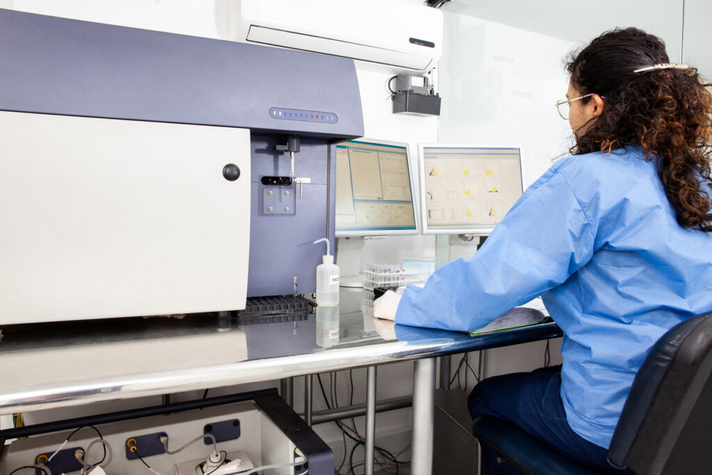
Fluorescence-activated cell sorting (FACS) is an advanced variant of flow cytometry that leverages fluorescent labels to sort and analyze cells, enabling researchers to isolate distinct populations with precision. FACS sorting is a crucial technique in various fields, such as hematopoiesis, oncology, and immunotherapy, allowing for detailed study of cellular functions and disease mechanisms. This method has become indispensable for scientists exploring the cellular underpinnings of health and disease. Read on to understand how FACS sorting works and its significant impact on modern research and clinical practices.
What does FACS stand for?
FACS stands for fluorescence-activated cell sorting, a specific type of flow cytometry. Other techniques, such as MACS (magnetic-activated cell sorting), use similar acronyms. By integrating FACS into their workflows, laboratories can accelerate discoveries, paving the way for groundbreaking advancements in medical and biological research.

How was FACS invented?
Fluorescence-activated cell sorting has a rich history that dates back to the 1960s. It was first developed by a team of geneticists at Stanford who created a technique combining flow cytometry principles and fluorescence microscopy to sort cells based on specific fluorescent markers. This groundbreaking work laid the foundation for modern cell sorting methods.
In the late 1970s, Dr. Leonard Herzenberg and his team at Stanford University significantly advanced the technology, introducing the first commercial FACS machine. Their innovations included the development of monoclonal antibodies, which could bind to specific cell surface markers and fluoresce, allowing for precise cell sorting. This innovation revolutionized immunology by enabling researchers to isolate and study distinct cell populations with unprecedented accuracy.
Over the years, FACS technology has continued to evolve, with advancements in laser technology, fluorescent dyes, and computing power. These improvements have expanded FACS’s capabilities, allowing for the simultaneous analysis of multiple parameters and enhancing the sensitivity and speed of cell sorting.
FACS has profoundly impacted biomedical research, playing a crucial role in fields such as cancer research, stem cell biology, and immunology. By enabling the precise isolation of specific cell types, FACS has facilitated numerous scientific breakthroughs, from identifying rare cell populations to developing targeted therapies.
The FACS Process
FACS protocols consist of four general phases:
- Sample preparation and labeling: Researchers begin by preparing a cell sample, ensuring cells are viable and suspended uniformly. Fluorescent labels—dyes attached to antibodies—are introduced to the sample, adhering to specific surface features unique to each cell type.
- Laser excitation and cell interrogation: Once labeled, cells are funneled through the flow cytometer one at a time. They encounter a laser beam that excites the fluorescent tags, causing each cell to emit light at varying wavelengths depending on their labels.
- Signal detection and system analysis: Sophisticated detectors within the FACS system capture the emitted light and the scattered light from cells. Forward scatter reveals information about cell size, while side light scatter provides insights into the granularity or internal complexity of the cells. The emitted fluorescence is indicative of the bound fluorescent tag, thus revealing the presence and quantity of specific cell markers.
- Cell sorting and collection: Post-detection, cells are electrically charged and pass through an electromagnetic field within the sorter. This field diverts cells into different containers based on their charge, effectively sorting them according to the predefined fluorescence profiles.
Once the samples are prepared, the rest of the process is automated, allowing for the sorting of thousands to millions of cells in a relatively short period. Scientists using FACS synergize precision optics, fluidics, and digital processing to deliver precise data on a cell-by-cell basis, making it a fundamental procedure in modern cellular analysis.
The Scientific Mechanisms of FACS
A handful of key scientific principles drive flow cytometry technology and the application of FACS.
Immunophenotyping and the Use of Antibodies
Immunophenotyping via flow cytometry and FACS is integral to cellular biology. It allows scientists to categorize cells based on the presence of specific cell surface proteins, also called markers. This process relies heavily on antibodies tagged with fluorescent labels that bind to these antigens.
Through binding, distinct cell types within complex mixtures can be identified and isolated. This level of specificity is essential to analyzing cells’ functional and phenotypic properties, such as in distinguishing different lymphocyte subsets within the immune system.
The Role of Fluorescence in Identifying Cell Populations
Fluorescence is at the heart of FACS, providing the means to detect and sort cells. Fluorophores attached to antibodies absorb light at one wavelength and emit it at another, acting as beacons that signal the presence of the cellular features they’re bound to. The flow cytometer’s lasers excite these fluorophores, and the emitted light is then captured, allowing for the characterization of each cell’s fluorescent profile.
The Dynamics of Single-Cell Sorting
Single-cell sorting is a delicate and precise process that requires the manipulation of individual cells based on their fluorescence profiles. After excitation and emission, droplets containing single cells are electrically charged. These charged droplets are then deflected into collection vessels by an electromagnetic field, enabling the isolation of single cells for downstream applications, such as single-cell genomics or cloning.
Data Analysis for Different Cell Characteristics
FACS sorts cells based on three main characteristics:
- Cell size (forward scatter): Also used in general flow cytometry, forward scatter correlates with cell size, providing an indirect measurement that helps distinguish between large and small cells.
- Granularity (side scatter): Also used in general flow cytometry, side scatter can detect elements of complexity within the cell, such as the presence of granules or the nucleus, offering insights into the cell’s internal structure.
- Fluorescence (emission): Also used in general flow cytometry and the cornerstone of FACS, the analysis of fluorescence intensity allows for the detection of specific cell populations. It can quantify the expression levels of surface receptors and other cellular features, providing valuable information on the cell’s identity and state.
Advantages and Limitations of FACS
Unlike general flow cytometry techniques, FACS is optimized for certain applications.
Advantages
The main advantage of FACS is its precise classification and analysis of cell subpopulations. This enables researchers to pinpoint and collect cells with high specificity, leading to more accurate results in studies of cellular behavior and function. Fluorescent labels allow for distinguishing even subtle differences in cell phenotypes.
Another major advantage is the method’s high-throughput capacity. Modern flow cytometers equipped with FACS technology can analyze and then sort thousands of cells per second, generating significant amounts of data in a relatively short time. This efficiency is vital in research fields where large sample sizes are essential for statistical significance and reliability.
The technology also enables simultaneous multiparametric analysis. This means that researchers can assess multiple characteristics of a single cell simultaneously—such as size, granularity, and the presence of various cell surface markers. This multi-dimensional analysis is crucial for comprehensive phenotyping, which is pivotal in understanding complex immune responses and diseases.
Disadvantages
The speed of FACS, while beneficial in high-throughput applications, can be a limiting factor. A balance must be struck between flow rate and accuracy, as too fast a rate can compromise the quality of cell sorting and data analysis.
Additionally, sample recovery rates can vary and are generally lower when sorting rare populations. As a positive selection process, FACS often requires starting with a larger number of cells. The process can also induce stress on cells, which may cause apoptosis and affect their viability for subsequent experiments.
Preparing samples can be time-consuming, requiring scientists to carefully stain cells with fluorescent antibodies—a precise and controlled procedure that ensures high specificity and prevents non-specific binding. Furthermore, the cost of FACS can be prohibitive. The equipment is expensive to purchase and maintain, and it requires trained personnel to operate. This significant investment can be a barrier for smaller laboratories or institutions with limited budgets.
FACS Research Applications
FACS analysis has created a niche across various fields of biological research due to its precision and efficiency in cell separation and analysis. Below are some of its major applications:
- Oncology tumor profiling: FACS enables the isolation of specific cancer cell populations for molecular profiling, which is crucial for understanding tumor progression, metastasis, and response to therapies. This facilitates the identification of biomarkers for diagnosis and the development of targeted treatments.
- Stem cell research: FACS allows scientists to isolate stem and progenitor cells from a mixed population based on cell surface markers. This aids in the study of their differentiation pathways and potential in regenerative medicine.
- Identifying immune cell subpopulations and apoptosis: FACS analysis identifies and sorts immune cell subpopulations, crucial for studying immune responses. It can sort T cell subtypes by intracellular cytokine staining and detect cellular apoptosis by identifying the molecules indicative of cell death, contributing to research on disease mechanisms and the development of immunotherapies.
- Vaccine and therapy development: By sorting and analyzing cells, FACS accelerates vaccine development, allowing scientists to effectively evaluate immune responses to various antigens. In therapeutic interventions, FACS-based phenotyping aids in patient stratification for personalized medicine, optimizing therapeutic outcomes.
Comparison With Other Cell Sorting Technologies
While FACS is a well-established cell sorting method, alternative technologies have emerged, each with its own set of advantages. Magnetic-activated cell sorting (MACS) utilizes magnetic particles to tag cells, allowing for separation using a magnetic field. Although MACS is generally less expensive and faster, it lacks the single-cell precision that FACS analysis offers.
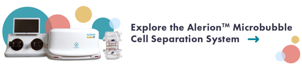
Another alternative is Akadeum’s buoyancy-based microbubble technology. Functionalized microbubbles bind target cells, allowing them to float to the surface for easy removal. This method is quicker and less equipment-intensive than FACS, offering a viable option for labs seeking efficient cell sorting without the need for expensive flow cytometers. Microbubbles are also far more gentle on cells, preserving their viability and function.
Each cell sorting technology has its place in the lab, with the choice often depending on the requirements of the specific study. Factors like the need for precision, speed, sample size, and budget constraints all play a role in determining whether general flow cytometry, FACS, MACS, microbubbles, or another method is the most suitable for a given application.
How Do Akadeum Microbubbles Compare to FACS Technology?
Fluorescence-activated cell sorting continues to be a transformative force in biomedical research, offering high specificity and versatility in cell analysis. As researchers and clinicians embrace the potential of personalized medicine and delve deeper into cellular mechanisms, the precision of FACS will remain invaluable.
However, innovations like Akadeum’s microbubble technology are poised to expand the accessibility and efficiency of cell sorting, heralding a new era of research where advanced technologies are within reach of every laboratory. No additional equipment is needed to use Akadeum’s microbubbles, reducing the cost of purchasing and storing magnets, columns, or other expensive materials.
Microbubble technology is easy to use and significantly reduces cell separation time. The entire process takes about 30 minutes for a typical negative selection kit or less than 10 minutes for Akadeum’s depletion kit. Microbubbles even isolate target cells at extremely high purity.
Additionally, microbubbles can be particularly beneficial when working with small sample sizes or when high cell viability is paramount. Our microbubble kits capture target cells, quickly and gently floating them to the surface of a biological sample for removal.
For those looking to integrate the latest in cell sorting efficiency and innovation, we invite you to explore how Akadeum’s groundbreaking microbubble technology can advance your research.
Contact us today to discover more about our solutions and how they can enhance your scientific endeavors.